
BIO-IMAGE ANALYSIS
The Necker Bio-image Analysis platform was created to meet the image analysis needs
of the Necker research campus. It specializes in bio-image processing and analysis services.
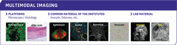
Its scope includes all imaging modalities from:
-
Different SFR platforms (such as microscopy and histology)
-
Resources common to the 2 institutes (Incucytes, Nanolive, etc.)
-
Systems specific to research teams on the Necker campus or outside.
All the work of the platform is done in scientific collaboration with the users
and the platforms concerned.
SERVICES
IMAGE ANALYSIS AS A SERVICE IS STRUCTURED AROUND 4 AXES
-
Advice: the Necker bio-image analysis platform is available for consultative meetings
at all stages of the project.
-
Analysis protocols: for each image analysis project: a first meeting will be planned
for a viewing of a few "raw" images to decide on the feasibility of the analysis.
In the event that the analysis platform cannot satisfy the desired analysis (lack of data, excessively altered image, etc.), an explanation of the disruptive analysis point(s)
will be provided and other strategies will be proposed.
-
Training: once the workflow has been established, the user will be trained
in the application of the analysis protocol and monitoring to support them in its use
will be systematically offered. At a minimum, a standard protocol will be rendered containing all the analysis steps from a few raw images. On request, a video tutorial
can be produced and provided to the team.
-
Results: Finally, the platform also supports if necessary in the interpretation of the data resulting from the analysis and offers its help on the technical points of the material and method of publication.
LIST OF SERVICES IN DETAIL
-
Advice and support on the possibilities in image analysis before the project,
during the project and after it with result discussion and help for paper and reviewing.
-
Training on the use of open-source (Fiji, Icy, Ilastik, QuPath, etc.) and licensed
(Imaris, etc.) analysis software.
-
Creating video tutorials for better laboratory knowledge transmission.
-
Design and drafting of specific analysis protocol.
-
Writing macros to automate your analysis and support in their use.
-
Script writing to compile your Excel results from the analysis and support for their reuse.
-
Provision of powerful analysis stations (remotely accessible) with AI capability adapted to analysis needs.
-
Development of new specific analysis approaches in collaboration with teams :
reuse of published plugin, Machine Learning and Deep Learning technics, etc.
-
Deep Learning 2D model creation.

EQUIPMENT
All open source softwares (FIJI, Ilastik, QuPath, Icy), are installed on all the system
of the image analysis Platform.
Artificial Intelligence (AI) neural network for 2D segmentation StarDist (FIJI/QuPath), Cellpose2 (FIJI QuPath) and SAM (QuPath), are installed on all GPU compatible workstations (AI).
FACULTÉ DE MEDECINE BUIDING
1st floor room 108b:
-
1 workstation with licenced software : Imaris latest version + AI GPU
(AMD 24core - 48threads / 128Go RAM / 8To storage / GPU Nvidia 8Go)
-
1 workstation dedicated to AI (with GPU) -> Début 2024
(Intel 12core - 24threads / 128Go RAM / 5To storage / GPU Nvidia 12Go)
-
1 analysis PC (Intel 6core - 12threads / 24Go RAM / 5To storage / GPU Ati 1Go)
IMAGINE BUIDING
2d floor (close to room 208):
-
1 workstation with licenced software : Imaris v9.5.3 + AI GPU
(Intel 20core - 40threads / 128Go RAM / 2x 2 storage / GPU Nvidia 8Go)
-
1 Analysis PC (Intel 4core - 8 threads / 32Go RAM / 2To storage / GPU Nvidia 2 Go)
EXAMPLES
The platform has a varied panel of analyses but it is also possible to develop,
in collaboration with the research teams, new approaches which are not yet included
in the portfolio.

Some examples below:
SEGMENTATION
Filaments like structure:
Primary cilia: Length and straightness measurements

Credit: Damelys Calderon
Spots like structure
number of spots per cell

Rossignol et al.- Neuropilin-1 cooperates with PD-1
in CD8 + T cells predicting outcomes in melanoma patients treated with anti-PD1 -
iScience 25,104353 June 17, 2022
Cell–like structure

Kaimal, Jay, & Thul, Peter. (2021). HPA Cell Image Segmentation Dataset (Version v2) [Data set]. Zenodo
Bacteria–like structure

Credit: omnipose GUI
Complex tissular structure
3D Nephron, cysts and vascular system if kidneys

T Blanc et al. - Three-dimensional architecture of nephrons in the normal and cystic kidney -
Kidney International (2021) 99, 632–645
COLOCALIZATION MEASUREMENT
By area (2D) and volume (3D) overlay

L. Cheradame et al. - STING protects breast cancer cells from intrinsic and genotoxic-induced DNA instability via a non-canonical, cell-autonomous pathway - Oncogene (2021) 40:6627 – 6640
DISTANCE ANALYSIS 3D/2D
Counting spots inside mitochondria

Credit: Eloïse Marques
Counting cells inside and at a certain distance of a delimitate tissular structure

Credit: Florence Vasseur
MEASUREMENTS
Volume / Area
Adipocytes area of a tissular structure

Ladraa et al. - PIK3CA gain-of-function mutation in adipose tissue induces metabolic reprogramming with Warburg-like effect and severe endocrine disruption - Sci. Adv. 8, eade7823 (2022)
Counting
number of foci per nuclei

L. Cheradame et al. - STING protects breast cancer cells from intrinsic and genotoxic-induced DNA instability via a non-canonical, cell-autonomous pathway - Oncogene (2021) 40:6627 – 6640
Intensity measurments

P. Renard et al. - Hydroxychloroquine sulfate: A novel treatment for lipin-1 deficiency? - Biomedicine & Pharmacotherapy 163 (2023) 114813
Morphological measurements

Bernheim et al. - Identification of Greb1l as a genetic determinant of crisscross heart in mice showing torsion
of the heart tube by shortage of progenitor cells- 2023, Developmental Cell 58, 2217–2234
OBJECT CLASSIFICATION
3D mitochondries segmentation and classification

E. Cosialls et al. - mTOR inhibition suppresses salinomycin-induced ferroptosis in breast cancer stem cells
by ironing out mitochondrial dysfunctions – Springer Nature Cell Death and Disease (2023) 14:744
TIME LAPSE ANALYSIS
Dynamic events
counting spots at different time point

I.Munoz et al. -Kinesin-1 regulates mast cell degranulation - JCB • Volume 215 • NumBer 2 • 2016
Tracking
measurements along time (speed, area, displacement, etc.)

Credit: Iwona Pranke
CREATION / IMPROVEMENT OF A 2D DEEPLEARNING MODEL

Credit: Thierry Capiaud
STAFF
Nicolas GOUDIN
Head
Pablo VARGAS
Scientific Referent
SCIENTIFIC STEERING COMMITTEE
IMAGINE
-
Sophie Thomas, PhD ; sophie.thomas@inserm.fr
-
Audrey Desgrange, PhD ; audrey.desgrange@institutimagine.org
INEM
-
Etienne Morel, PhD ; etienne.morel@inserm.fr
-
Julien Diana, PhD ; julien.diana@inserm.fr
FEES
For any pricing indication, please contact the platform at this address : nicolas.goudin@inserm.fr
PUBLICATIONS
Mitochondrial dynamics and metabolic regulation control T cell fate in the thymus.
Elhage R, Kelly M, Goudin N, Megret J, Legrand A, Nemazanyy I, Patitucci C, Quellec V, Wai T, Hamaï A, Ezine S. Front Immunol. 2024 Jan 15;14:1270268. doi: 10.3389/fimmu.2023.1270268. eCollection 2023. PMID: 38288115
The AMPK-Sirtuin 1-YAP axis is regulated by fluid flow intensity and controls autophagy flux in kidney epithelial cells.
Claude-Taupin A, Isnard P, Bagattin A, Kuperwasser N, Roccio F, Ruscica B, Goudin N, Garfa-Traoré M, Regnier A, Turinsky L, Burtin M, Foretz M, Pontoglio M, Morel E, Viollet B, Terzi F, Codogno P, Dupont N. Nat Commun. 2023 Dec 5;14(1):8056. doi: 10.1038/s41467-023-43775-1. PMID: 38052799
Hemifacial myohyperplasia is due to somatic muscular PIK3CA gain-of-function mutations and responds to pharmacological inhibition.
Bayard C, Segna E, Taverne M, Fraissenon A, Hennocq Q, Periou B, Zerbib L, Ladraa S, Chapelle C, Hoguin C, Kaltenbach S, Villarese P, Asnafi V, Broissand C, Nemazanyy I, Autret G, Goudin N, Legendre C, Authier FJ, Viel T, Tavitian B, Gitiaux C, Fraitag S, Duong JP, Delcros C, Sergent B, Picard A, Dussiot M, Guibaud L, Khonsari R, Canaud G. J Exp Med. 2023 Nov 6;220(11):e20230926. doi: 10.1084/jem.20230926. Epub 2023 Sep 15. PMID: 37712948
Hydroxychloroquine sulfate: A novel treatment for lipin-1 deficiency?
Renard P, Caccavelli L, Legendre A, Tuchmann-Durand C, Balakirouchenane D, Blanchet B, Narjoz C, Straube M, Hubas A, Garros A, Mention K, Bednarek N, Goudin N, Broissand C, Schlatter J, Cisternino S, Cagnard N, van Endert P, Diana J, de Calbiac H, de Lonlay P. Biomed Pharmacother. 2023 Jul;163:114813. doi: 10.1016/j.biopha.2023.114813. Epub 2023 May 5. PMID: 37150031
DOCK11 deficiency in patients with X-linked actinopathy and autoimmunity.
Boussard C, Delage L, Gajardo T, Kauskot A, Batignes M, Goudin N, Stolzenberg MC, Brunaud C, Panikulam P, Riller Q, Moya-Nilges M, Solarz J, Repérant C, Durel B, Bordet JC, Pellé O, Lebreton C, Magérus A, Pirabakaran V, Vargas P, Dupichaud S, Jeanpierre M, Vinit A, Zarhrate M, Masson C, Aladjidi N, Arkwright PD, Bader-Meunier B, Baron Joly S, Benadiba J, Bernard E, Berrebi D, Bodemer C, Castelle M, Charbit-Henrion F, Chbihi M, Debray A, Drabent P, Fraitag S, Hié M, Landman-Parker J, Lhermitte L, Moshous D, Rohrlich P, Ruemmele F, Welfringer-Morin A, Tusseau M, Belot A, Cerf-Bensussan N, Roelens M, Picard C, Neven B, Fischer A, Callebaut I, Ménager M, Sepulveda FE, Adam F, Rieux-Laucat F. Blood. 2023 Jun 1;141(22):2713-2726. doi: 10.1182/blood.2022018486. PMID: 36952639
PIK3CA gain-of-function mutation in adipose tissue induces metabolic reprogramming with Warburg-like effect and severe endocrine disruption.
Ladraa S, Zerbib L, Bayard C, Fraissenon A, Venot Q, Morin G, Garneau AP, Isnard P, Chapelle C, Hoguin C, Fraitag S, Duong JP, Guibaud L, Besançon A, Kaltenbach S, Villarese P, Asnafi V, Broissand C, Goudin N, Dussiot M, Nemazanyy I, Viel T, Autret G, Cruciani-Guglielmacci C, Denom J, Bruneau J, Tavitian B, Legendre C, Dairou J, Lacorte JM, Levy P, Pende M, Polak M, Canaud G. Sci Adv. 2022 Dec 9;8(49):eade7823. doi: 10.1126/sciadv.ade7823. Epub 2022 Dec 9. PMID: 36490341
Neuropilin-1 cooperates with PD-1 in CD8+ T cells predicting outcomes in melanoma patients treated with anti-PD1.
Rossignol J, Belaid Z, Fouquet G, Guillem F, Rignault R, Milpied P, Renand A, Coman T, D'Aveni M, Dussiot M, Colin E, Levy J, Carvalho C, Goudin N, Cagnard N, Côté F, Babdor J, Bhukhai K, Polivka L, Bigorgne AE, Halse H, Marabelle A, Mouraud S, Lepelletier Y, Maciel TT, Rubio MT, Heron D, Robert C, Girault I, Lebeherec D, Scoazec JY, Moura I, Condon L, Weimershaus M, Pages F, Davoust J, Gross D, Hermine O. iScience. 2022 May 5;25(6):104353. doi: 10.1016/j.isci.2022.104353. eCollection 2022 Jun 17. PMID: 35874918
The critical role of the TB5 domain of fibrillin-1 in endochondral ossification.
Delhon L, Mougin Z, Jonquet J, Bibimbou A, Dubail J, Bou-Chaaya C, Goudin N, Le Goff W, Boileau C, Cormier-Daire V, Le Goff C. Hum Mol Genet. 2022 Nov 10;31(22):3777-3788. doi: 10.1093/hmg/ddac131. PMID: 3566086
2D and 3D Human Induced Pluripotent Stem Cell-Based Models to Dissect Primary Cilium Involvement during Neocortical Development.
Boutaud L, Michael M, Banal C, Calderon D, Farcy S, Pernelle J, Goudin N, Maillard C, Dimartino C, Deleschaux C, Dupichaud S, Lebreton C, Saunier S, Attié-Bitach T, Bahi-Buisson N, Lefort N, Thomas S. J Vis Exp. 2022 Mar 25;(181). doi: 10.3791/62667. PMID: 35389978
STING protects breast cancer cells from intrinsic and genotoxic-induced DNA instability via a non-canonical, cell-autonomous pathway.
Cheradame L, Guerrera IC, Gaston J, Schmitt A, Jung V, Goudin N, Pouillard M, Radosevic-Robin N, Modesti M, Judde JG, Cairo S, Goffin V. Oncogene. 2021 Dec;40(49):6627-6640. doi: 10.1038/s41388-021-02037-4. Epub 2021 Oct 8. PMID: 34625708